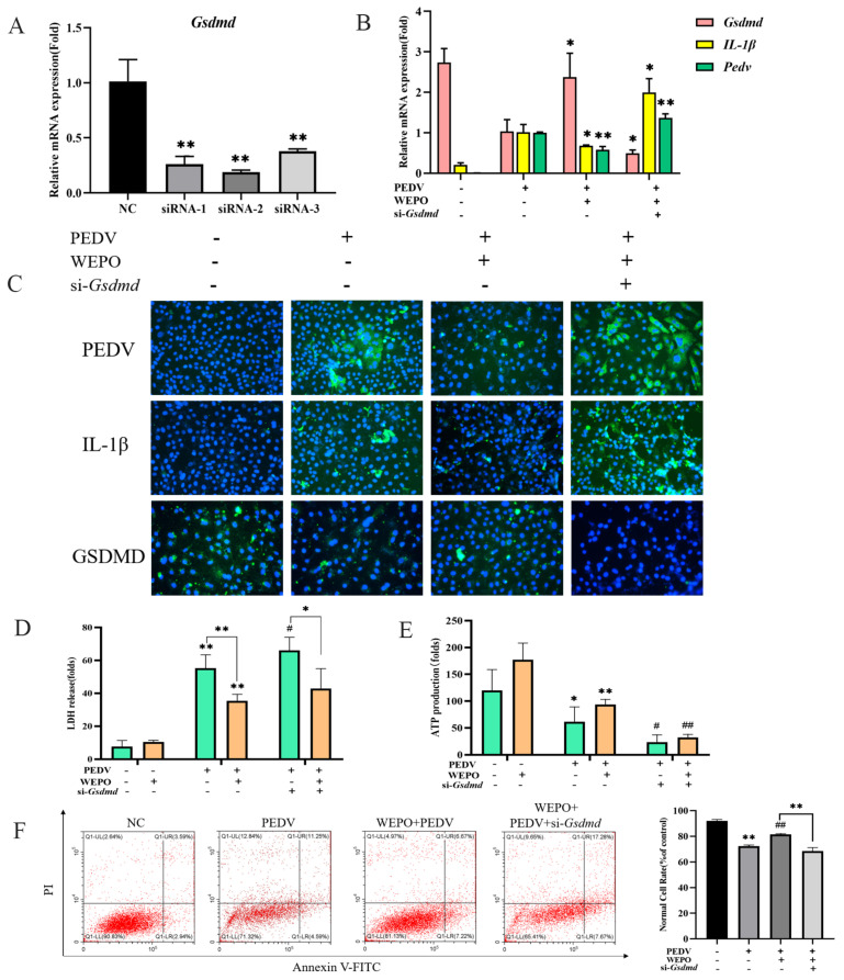
Figure 4.
GSDMD participates in the pyroptosis of WEPO-inhibited PEDV-infected Vero cells. (A) Detection of the interference efficiency of siGSDMD with RT-PCR. The data are expressed as the means ± SD (n = 3). ** p < 0.01 vs. control. (B) GSDMD was knocked out, and IL-1β, GSDMD, and PEDV expression was detected through RT-PCR. The data are expressed as the means ± SD (n = 3). * p < 0.05 and ** p < 0.01 vs. positive control (PEDV + WEPO). (C) Expression of PEDV and IL-1β after GSDMD knockout using an immunofluorescence assay. (The magnification of a microscope is 200 times). (D) Detection of LDH release after adding PEDV followed by adding purslane and knocking off GSDMD. The data are expressed as the means ± SD (n = 3). * p < 0.05. ** p < 0.01 vs. control. # p < 0.05 vs. positive control (PEDV). (E) Detection of ATP generation after adding PEDV followed by adding purslane and knocking off GSDMD. The data are expressed as the means ± SD (n = 3). * p < 0.05 and ** p < 0.01 vs. control. # p < 0.05 and ## p < 0.01 vs. positive control (PEDV). (F) Detection of apoptosis in Vero cells after knocking out GSDMD with flow cytometry. ** p < 0.01 vs. control. ## p < 0.01 vs. positive control (PEDV).

