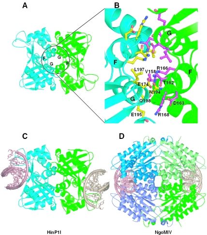
Figure 5.
Potential link between the dimeric form of HinP1I and the tetrameric restriction enzymes (A) The HinP1I dimer interface mediated by a crystallographic 2-fold symmetry. (B) The enlarged dimer interface of HinP1I consists of residues from helices αG and αF. (C) A model of HinP1I dimer docked with two DNA molecules. (D) Structure of a tetramer of the NgoMIV restriction endonuclease in complex with two DNA molecules (PDB 1FIU). Two primary dimers (blue and green) are positioned back-to-back to each other.

