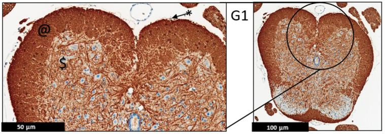
Figure 6.
Representative histology of the spinal cords in EAE mice. G1: Cross-section from MBP immunohistochemistry spinal cord staining. Panoramic view of normal spinal cord stained with myelin basic protein. Again, there is well demarcation between white (@) and gray ($) matter; intact thin leptomeninges (*). The staining is uniform in white matter, (n = 3).

