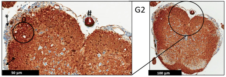
Figure 7.
Representative histology of the spinal cords in EAE mice. G2: Cross-section from MBP immunohistochemistry spinal cord staining. EAE mice received a daily IP injection of scramble peptide (G2) until day 21 post-EAE induction. Leptomeninges (*) are thickened by lymphocytic infiltrates, which appear to extend to the adjacent white matter. There is no significant loss of myelin basic protein staining in white matter (properly heavy staining—note, there is non-specific staining in endothelial cells of some blood vessels (#)). Patchy vacuolation (O) is evident, (n = 3).

