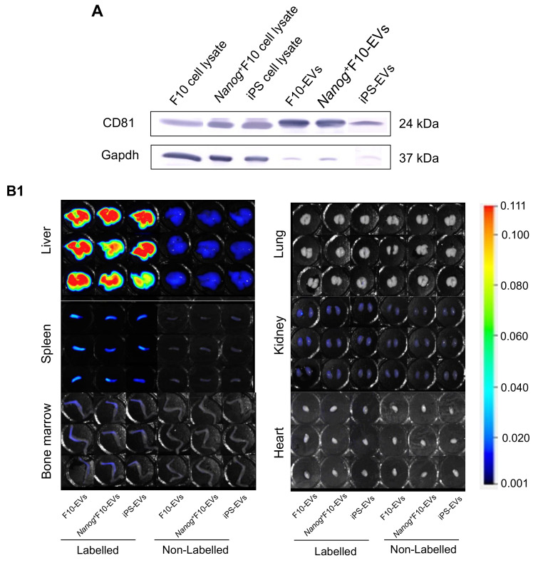
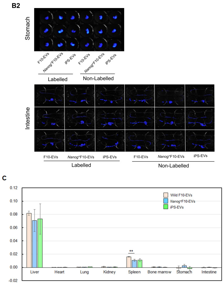
Figure 1.
Accumulation of EVs in eight organs. (A) Confirmation of CD81-positive fractions of EVs. (B1) Imaging of F10 EVs, Nanog+F10-EVs, and iPS EVs in liver, lung, spleen, kidney, bone marrow, and heart. EVs were labeled with CellVue™ NIR815 or unlabeled. n = 3 mice per each of six con-ditions. (B2) Same as (B1) for stomach and intestine. (C) Comparison of the accumulated densities of EVs in different organs. Mean ± SD, n = 3, ** p < 0.01.


