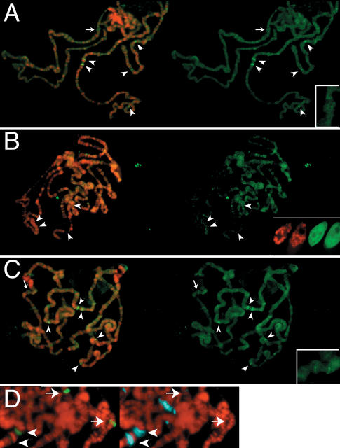
Figure 4.
Histone replacement uses newly produced H3.3. Late-third-instar larvae containing a heat-shock-inducible histone-GFP construct were heat-shocked for 1 h to induce the construct and endogenous HSP genes, and then allowed to recover for 4 h. (A–C) DNA (red) and histone-GFP (green) images are shown as merges on the left, and the histone-GFP signal alone are shown on the right. (A) Full-length H3.3-GFP has deposited specifically at the heat-shock loci (arrowheads indicate the 87A, 87C, 95D, 63E, and 67F loci). (B) H3-GFP is not incorporated into chromatin. H3-GFP fills the interchromatin space within unfixed salivary gland nuclei after induction, but remains diffuse (inset). (C) Spreads with H3.3-GFP and two HSP70 promoter transgene arrays show intense H3-GFP signals at HSP loci but little signal at the arrays. Arrows in A and C indicate the 43E region without an array (A) and with an array (C). (Insets) Magnified images of 43E are shown. (D) Chromosomes from larvae carrying P[H3.3-GFP]B6 and the HSP70 promoter transgene arrays at positions 30A and 43E during a 20-min heat-shock induction. The endogenous HSP70 loci (arrowheads) bind moderate amounts of HSF activator (green), while arrays (arrows) bind large amounts. Conversely, HSP70 loci have large amounts of engaged RNA polymerase II (blue), while arrays have little.

