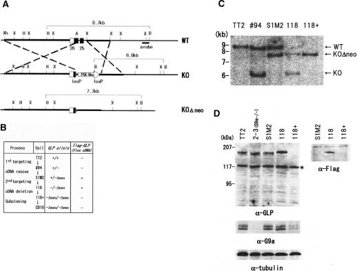
Figure 2.
Generation of GLP-deficient mice and ES cells. (A) Partial restriction maps of the mouse GLP locus (exons 25 and 26, top), and the targeted GLP locus containing a neomycin cassette (middle) are shown. (Bottom) The neomycin cassette can be excised from the targeted locus for disruption of the second allele. The portions of the endogenous locus that were used for constructing the targeting plasmid are shown as thick lines. (Xh) XhoI; (H) HindIII; (X) XbaI; (A) ArtII; (B) BamHI. (B) Flow diagram for the establishment of GLP-deficient ES cells. To disrupt GLP conditionally with Cre-recombinase, loxP-flanked (flox) Flag-GLP-cDNA was introduced before the second GLP targeting. The 118 cells, which expressed only exogenous Flag-GLP-cDNA, were treated with 5′OHT to excise the Flag-GLP-cDNA. The resultant cells (118+) successfully carried a GLP-null genotype. (C) Southern blot analyses of GLP alleles described in A. Disruption of the GLP locus was confirmed by Southern blot analysis using BamHI/HindIII-digested DNA probed with a genomic portion located outside of the targeting construct as shown in A.(D) GLP deficiency was confirmed by Western blot analyses using total lysates corresponding to 105 cells. Endogenous GLP protein was detected at a molecular weight of ∼170 kDa. (Top) Asterisks represent nonspecific signals detected by the secondary antibodies. (Bottom) Tubulin contents were also determined as a loading control.

