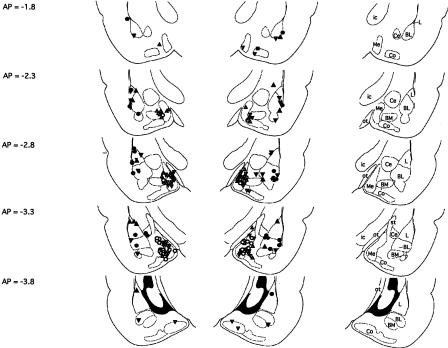
Figure 1.
Cannula tip placements transcribed onto atlas plates adapted from Paxinos and Watson (1997). Placements for rats included in the primary analyses of Experiments 1A, 1B, and 2 (basolateral amygdala) are indicated by filled circles, filled triangles (point up), and filled triangles (point down), respectively. Placements for rats included in the primary analyses of Experiments 3, 4A, and 4B (medial amygdala) are indicated by open circles, open triangles (point up), and open triangles (point down), respectively. The distance from bregma is indicated to the left; nuclei and major fiber bundles within the plane of section are identified to the right. BL, basolateral amygdaloid nucleus; BM, basomedial amygdaloid nucleus; Ce, central amygdaloid nucleus; Co, cortical amygdaloid nucleus; ic, internal capsule; L, lateral amygdaloid nucleus; Me, medial amygdaloid nucleus; ot, optic tract; st, stria terminalis.

