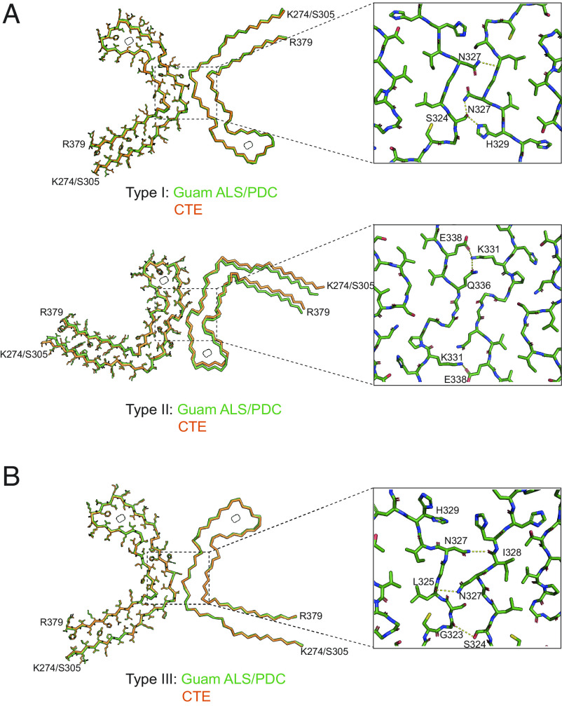
Fig. 2.
Comparison of tau filaments from Guam ALS/PDC and CTE. (A) Overlays of CTE Type I filaments (Top) and CTE Type II filaments (Bottom) from Guam ALS/PDC (green) and CTE (orange), with one protofilament shown in all atoms and the other protofilament shown as backbone. Insets: Zoomed-in views of the protofilament interfaces of both types of filaments. (B) Overlay of CTE Type III filaments from Guam ALS/PDC (green) and CTE (orange), shown as in (A). Inset: Zoomed-in view of the protofilament interface of CTE Type III filaments.

