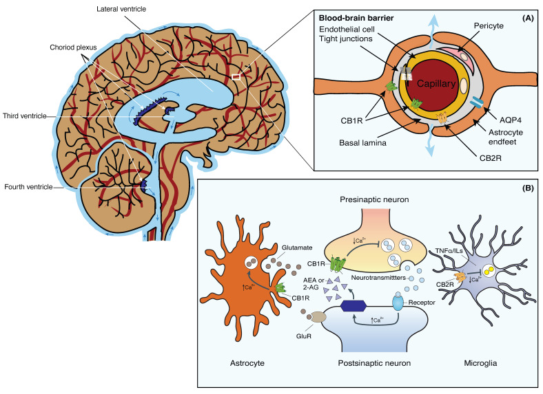
Figure 1.
Schematic representation of the brain’s fluid compartments and endocannabinoid receptors in the nervous and glia system. Main: The cerebral ventricles are a system of interconnected, fluid-filled cavities within the brain that contribute to cerebrospinal fluid (CSF) production and circulation. The four ventricles include the paired lateral ventricles, the third ventricle, and the fourth ventricle, connected by the cerebral aqueduct. These ventricles are lined with ependymal cells and house the choroid plexus, a specialized structure responsible for CSF secretion. Inset (A): Tight junctions between the blood endothelial cells constitute the BBB, restricting macromolecules from moving freely from the blood into the brain parenchyma. Fluid and solutes diffuse into the brain parenchyma from the perivascular space located between endothelial cells and astrocytic endfeet that expresses AQP4 and CB1R. CB1Rs are primarily located on the luminal side of the BBB endothelium. CB2Rs, on the other hand, are located on the abluminal side of the BBB. Inset (B): 2-AG or AEA are synthesized from phospholipids on demand. Activation of presynaptic CB1R negatively modulates cell calcium influx and the release of neurotransmitters in neurons. Stimulation of CB1R in astrocytes positively modulates calcium influx and glutamate release. Activation of CB2R in microglia negatively affects the release of TNFα and ILs. Abbreviations: AQP4, Aquaporin-4 water channel; BBB, blood-brain barrier; CB1R, Cannabinoid receptor 1; CB2R, Cannabinoid receptor 2; CSF, cerebrospinal fluid; 2-AG, 2-acylglycerol; AEA, anandamide; GluR, glutamate receptors; ILs, interleukins; TNFα, tumor necrosis factor-α. This figure was made in Adobe Illustrator 2020, modified from Jessen et al., 2015 [3].

