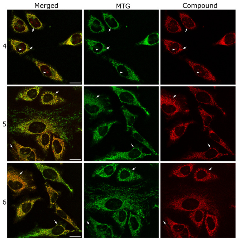
Figure 4.
Colocalization studies of COUPY dyes 4 (top), 5 (middle), and 6 (bottom) with MitoTracker Green FM (MTG). Single confocal planes of HeLa cells incubated with the corresponding coumarin dye (2 μM, λEx = 561 nm, λEm = 575–650 nm, red) and MTG (1 μM, λEx = 488 nm, λEm = 500–550 nm, green) both for 30 min at 37 °C. Left: merged images; center: MTG signal; right: compound signal. White arrows point out some colocalizing mitochondria, and white arrowheads in the top row point out nucleoli. Scale bar: 20 μm; all images are at the same scale.

