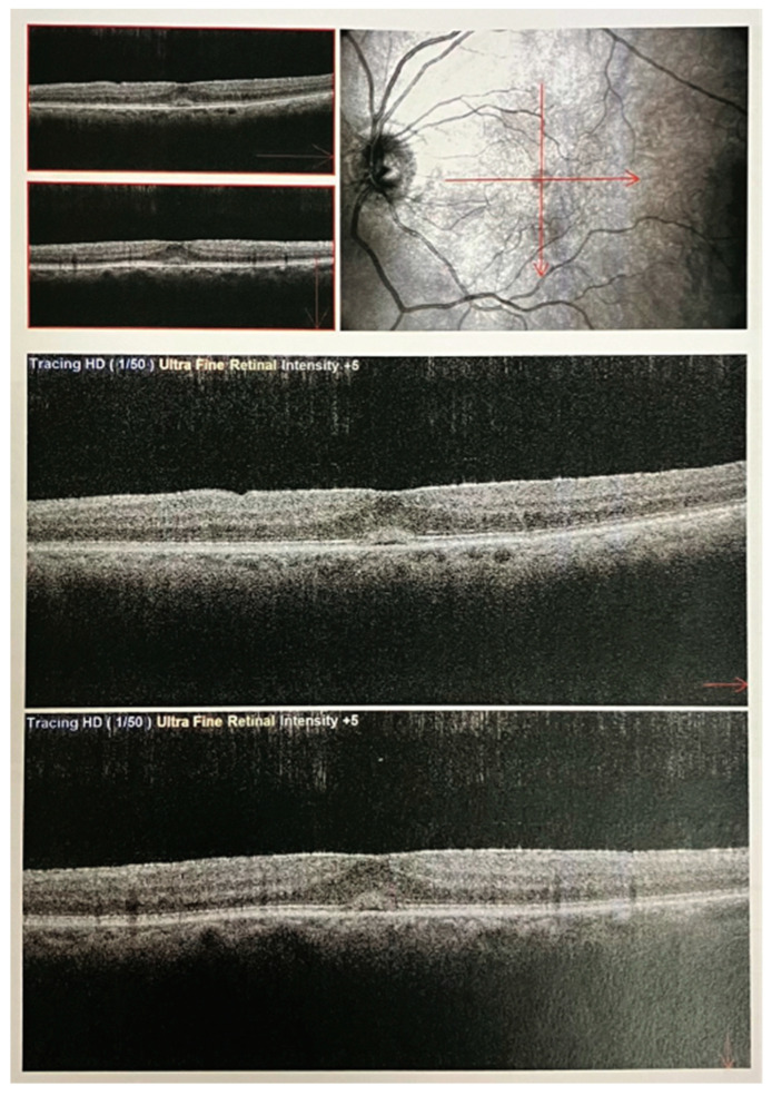
Figure 4.
OCT macular scans in a patient with tractional pucker and linearization. SD-OCT images using a Nidek Optical Coherence Tomography (OCT) RS-3000 instrument. The OCT macular scans (at the specific position indicated with the red lines) illustrate the macular microstructure in a patient diagnosed with tractional pucker and linearization. The image captures detailed cross-sectional views of the macula, highlighting the pathological changes associated with tractional forces on the retinal surface. Notably, the scans reveal the presence of epiretinal membranes causing distortion and traction on the retinal layers, leading to the linearization of the macular architecture. The hyper-reflective bands represent the fibrous tissue causing the tractional forces, while disruptions in the normal retinal layers underscore the impact of this condition on retinal morphology.

