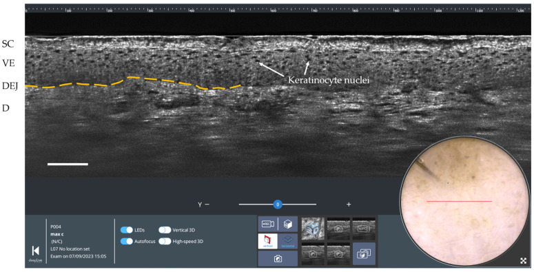
Figure 3.
Vertical LC-OCT and dermoscopy image of healthy skin using deepLive™ software. Scale bar: 100 µm. The red horizontal line superimposed on the dermoscopic image delineates the location of the vertical sectional LC-OCT image. SC: stratum corneum; VE: viable epidermis; DEJ: dermal-epidermal junction (partially marked by the yellow dashed line); D: dermis.

