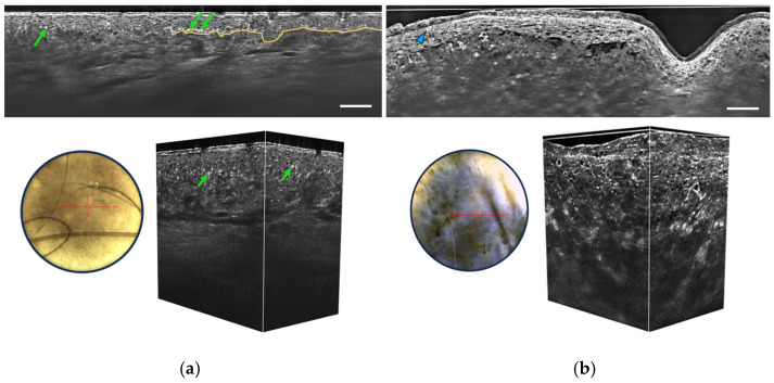
Figure 6.
Vertical sectional LC-OCT image, dermoscopic image (left), and 3D LC-OCT reconstruction (right) of a junctional nevus (a) and a melanoma (b). The red horizontal line superimposed on the dermoscopic images delineates the location of the sectional LC-OCT images. (a) Regular organization of keratinocytes in the viable epidermis, pigmentation appearing as white discs (green arrows), well-defined DEJ partially marked by the yellow line. (b) Disorganized epidermis, pagetoid cells (blue arrow), atypical cells in the dermis, and damaged DEJ. Scale bars: 100 µm.

