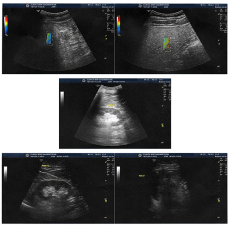
Figure 3.
Images show liver with normal topography, shape, dimensions, contours and surface. Homogeneous liver parenchyma echotexture, with signs of mild fatty infiltration, without focal lesions. Elastography with the shear wave method (Logiq S8) showing mean stiffness (Kpa) of 5.70 corresponding to non-significant fibrosis (FO-F1) on the METAVIR scale.

