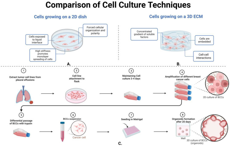
Figure 5.
There are key differences between the utilization of 2D cell cultures and 3D cell cultures. Part (A) shows that breast cancer cells grown on a 2D dish have a forced cellular organization and polarity, high stiffness to promote monolayer of spreading cells, and no cell–cell interaction. Part (B) illustrates 3D cell culturing techniques to embed breast cancer cells in ECM, incorporate cell–cell interactions, and form a more complex culturing system. Part (C) illustrates most breast cancer cell line derivation from pleural effusions to plating in a 2D culture flask for amplification of the differentiated cells. While organoid formation can then be formed after proper breast cancer proliferation and embedding in the proper ECM material, see Section 3.3.2 for more information.

