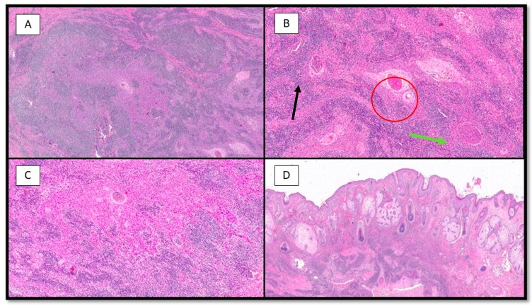
Figure 3.
(A) Photomicrograph showing a neoplastic lesion that is difficult to discern at this magnification into its two components: epithelial and inflammatory (hematoxylin–eosin, 10×). (B) Histopathological micrograph showing the two different components of the lesion: large, epithelioid cells with palely eosinophilic cytoplasm (green arrow) and brisk lymphoplasmocytic inflammatory infiltrate (black arrow). Note the important presence of squamous differentiation (red circle) (hematoxylin–eosin, 20×). (C) Low-power magnification of B (hematoxylin–eosin, 20×). (D) Photomicrograph showing no connection of the neoplasm with the overlying epidermis (H&E, 4×).

