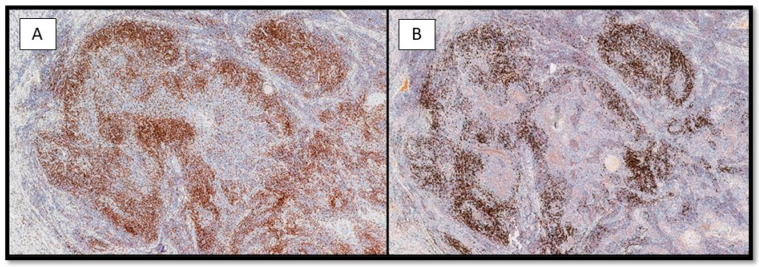
Figure 6.
(A) Histological photomicrograph showing a rich lymphocytic inflammatory infiltrate composed of CD3-T cells, surrounding the epithelial strands of the neoplasm (immunohistochemistry for CD3, 10×). (B) Photomicrograph showing CD20-B cells, surrounding the epithelial strands of the neoplasm in a similar way of the CD3-T cells (immunohistochemistry for CD20, 10×).

