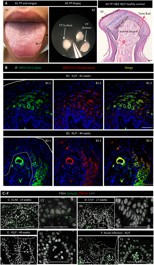
Figure 1. Severe Acute Respiratory Syndrome Coronavirus 2 Persists in Fungiform Papillae from Postacute Sequelae of Covid-19 Patients.
Panel A1 shows a human tongue with a fungiform papilla outlined in a black circle. Panel A2 shows excised fungiform papillae (FP) under a dissection microscope. Panel A3 shows hematoxylin and eosin (H&E) staining of an fungiform papilla. The black dotted line delineates the epithelial layer from the lamina propria, whereas the blue dotted line delineates the epithelial suprabasal from basal cell layers. The red dotted line outlines a taste bud. The surface of the fungiform papilla is covered by a keratinized cell layer. Panel B presents representative images of immunofluorescent staining for severe acute respiratory syndrome coronavirus 2 (SARS-CoV-2) spike (green) and nucleocapsid (red) proteins in FP epithelium from two patients experiencing chronic taste disturbances post–SARS-CoV-2. Nuclei are stained in blue using 4′,6-diamidino-2-phenylindole (DAPI). The solid white line indicates the surface of FP, and the white dotted line demarcates the epithelium from lamina propria. Note consistent colocalization of spike (S) and nucleocapsid proteins, both of which are present in the FP epithelium and in a blood vessel in the lamina propria in Panel B2. Panels C to F, fluorescence in situ hybridization (FISH), demonstrate the presence of viral RNA using a probe for S (green dots) and a probe for the SARS-CoV-2 open reading frame 1 ab (ORF1ab) negative-strand RNA, produced when the virus is replicating (red dots). Inserts show a higher magnification of the areas marked by rectangles. In all instances, note the presence of S. The presence of ORF1ab indicates replication of the virus in FP. Scale bars indicate 50 μm. F denotes female; and M, male.

