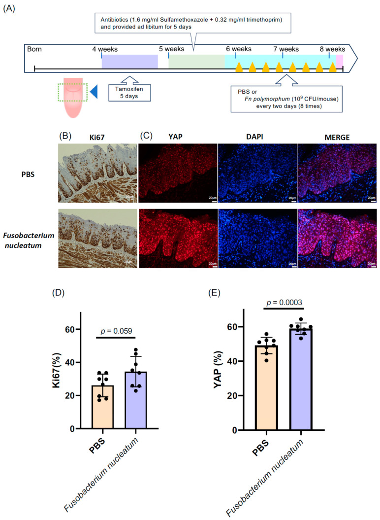
Figure 4.
Fusobacterium nucleatum isolated from the oral cavity of oral cancer patients was applied to the tongues of epithelial cell-specific Mob1a/b DKO mice (tgMob1DKO). (A) Experimental scheme for F. nucleatum-treated tgMob1DKO mice. Tamoxifen was brushed daily for 5 days onto the tongues of 4-week-old tgMob1DKO mice. The application area of tamoxifen is illustrated in the dotted square. Mice received water containing antibiotics, sulfamethoxazole, and trimethoprim, for 5 days starting from 5 weeks old. Subsequently, mice were orally treated eight times with F. nucleatum polymorphum (n = 8) or PBS (n = 8) every two days post antibiotic administration. Mice were euthanized post treatment, and their tongue tissues were analyzed histologically. (B) Representative Ki67 immunostaining of tongue epithelium from mice in (A). Scale bar: 20 μm. (C) Representative images showcasing the immunofluorescent detection of YAP (red) in tongue epithelium from mice in (A). DAPI (blue) stains nuclei. Scale bar: 20 μm. (D) Percentages of Ki67-positive cells from sections in (B) are shown in bar plots. (E) Percentages of YAP-positive cells from sections in (C) are represented in bar plots. Data are presented as means ± SEM with Student’s t-test applied.

