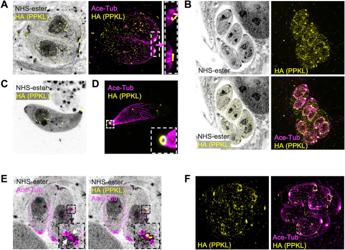
Fig 2.
Ultrastructure expansion microscopy reveals PPKL in basal and apical structures. Intracellular and extracellular parasites were fixed with paraformaldehyde, expanded in acrylamide gels, and stained with NHS-ester, anti-HA, and anti-acetylated tubulin. Images were captured by LSM 900 with Airyscan. (A) Image of intracellular non-dividing parasites. The white box frames the region expanded to the right of the arrow. (B) Images of intracellular dividing parasites. (C and D) Images of extracellular parasites. The white framed zone in panel D is zoomed in and shown in the lower right corner. (E) Images of intracellular parasites. The parasite on the right has started daughter parasite assembly, as shown by duplicated centrosomes, preconoidal regions (yellow dots labeled by PPKL), and apical polar rings (indicated with white arrows), which are framed in a black box. The box is enlarged to the left showing the acetylated tubulin signal and to the right showing both anti-acetylated tubulin and anti-HA. (F) Images show intracellular parasites in a late stage of division.

