Fig 1.
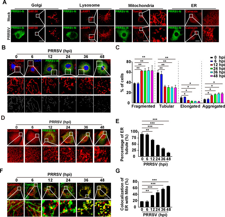
PRRSV infection induces the morphological alterations of mitochondria and ER, and ER-mitochondria contacts. (A) MARC-145 cells were mock-infected or infected with PRRSV (MOI = 1) for 24 h. The morphologies of Golgi (GM130), lysosomes (LAMP1), mitochondria (Tom20), and ER (calnexin) were monitored by immunofluorescence analysis. Scale bar: 10 µM. (B) MARC-145 cells were infected with PRRSV (MOI = 1) for 0–48 h. The morphology of mitochondria (Tom20) was monitored by immunofluorescence analysis. Scale bar: 10 µM. (C) Quantification of the fragmented, tubular, elongated, and aggregated mitochondria from (B) (n = 30). *P < 0.05, **P < 0.01. (D) MARC-145 cells were infected with PRRSV (MOI = 1) for 0–48 h. The morphology of ER (calnexin) was monitored by immunofluorescence analysis. Scale bar: 10 µm. (E) Quantification of the percentage of ER tubule from (D) (n = 30). **P < 0.01, ***P < 0.001. (F) MARC-145 cells were infected with PRRSV (MOI = 1) for 0–48 h. The co-localization of ER (calnexin) with mitochondria (Tom20) was monitored by immunofluorescence analysis. Scale bar: 10 µm. (G) Quantification of the co-localization of ER with mitochondria from (F) (n = 30). **P < 0.01, ***P < 0.001.

