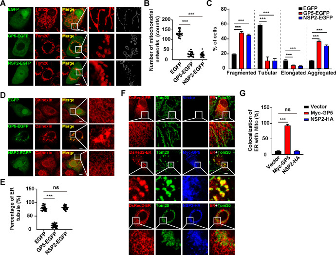
Fig 2.
PRRSV GP5 is responsible for ER-mitochondria contact. (A) MARC-145 cells were transfected with EGFP, GP5-EGFP, or NSP2-EGFP plasmids for 24 h. The morphology of mitochondria (Tom20) was monitored by immunofluorescence analysis. Scale bar: 10 µm. (B) Quantification of mitochondrial networks number from (A) (n = 30). ***P < 0.001. (C) Quantification of the fragmented, tubular, elongated, and aggregated mitochondria from (A) (n = 30). ***P < 0.001. (D) MARC-145 cells were transfected with EGFP, GP5-EGFP, or NSP2-EGFP plasmids for 24 h. The morphology of ER (calnexin) was monitored by immunofluorescence analysis. Scale bar: 10 µm. (E) Quantification of the percentage of ER tubules from (D) (n = 30). ***P < 0.001. ns, no significance. (F) MARC-145 cells were co-transfected with pDsRed2-ER and vector, Myc-GP5, or NSP2-HA for 24 h. The co-localization of ER (DsRed) with mitochondria (Tom20) was monitored by immunofluorescence analysis. Scale bar: 10 µm. (G) Quantification of the co-localization of ER with mitochondria from (F) (n = 30). ***P < 0.001. ns, no significance.

