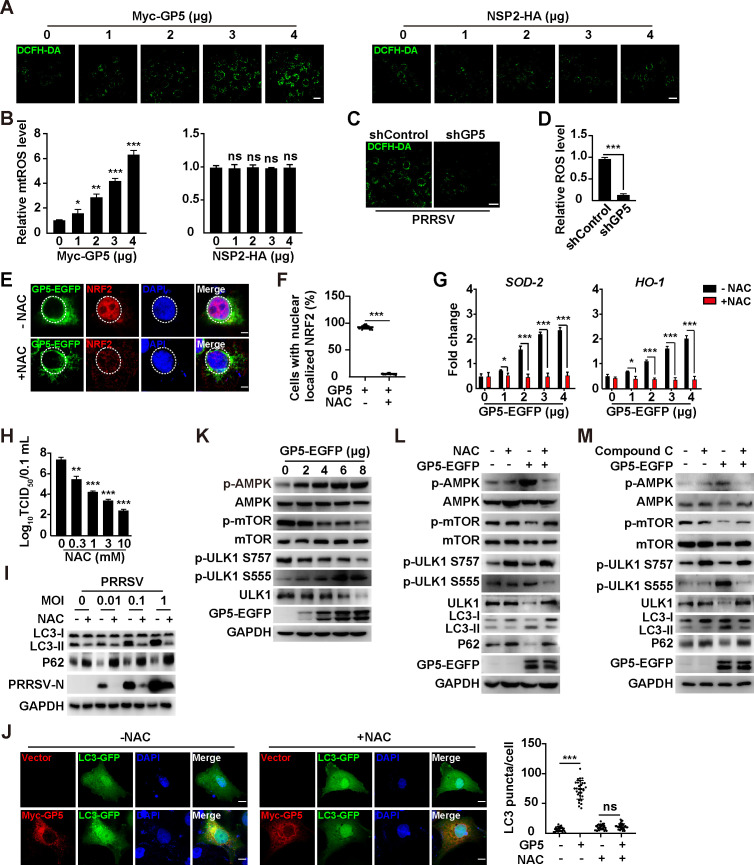
Fig 5.
PRRSV GP5 induces ROS and activates autophagy through the AMPK/mTOR/ULK1 axis. (A) MARC-145 cells were transfected with GP5-EGFP (0–4 µg) or NSP2-HA (0–4 µg) for 24 h. Intracellular ROS was determined by DCFH-DA staining. Scale bar: 10 µm. (B) Quantification of the relative ROS levels from (A). *P < 0.05, **P < 0.01, ***P < 0.001. ns, no significance. (C) shControl and shGP5 MARC-145 cells were infected with PRRSV (MOI = 1) for 24 h. Intracellular ROS was determined by DCFH-DA staining. Scale bar: 10 µm. (D) Quantification of the relative ROS levels from (C). ***P < 0.001. (E) MARC-145 cells were transfected with GP5-EGFP (4 µg) and simultaneously treated with vehicle or NAC (10 mM) for 24 h. The subcellular localization of GP5-EGFP and NRF2 was detected by immunofluorescence analysis. Scale bar: 10 µm. (F) Quantification of cells with nuclear-localized NRF2 from (E). ***P < 0.001. (G) MARC-145 cells were transfected with GP5-EGFP (0–4 µg) and simultaneously treated with vehicle or NAC (10 mM) for 24 h. The mRNA levels of SOD-2 and HO-1 were analyzed by qRT-PCR analysis. *P < 0.05, ***P < 0.001. (H) MARC-145 cells were infected with PRRSV (MOI = 1) and simultaneously treated with NAC (0–10 mM) for 48 h. Viral titers were assessed by TCID50 assay. **P < 0.01, ***P < 0.001. (I) MARC-145 cells were infected with PRRSV (MOI = 0–1) and treated with NAC (10 mM) as indicated for 48 h. LC3-I, LC3-II, P62, PRRSV-N, and GAPDH were analyzed by immunoblotting analysis. (J) MARC-145 cells were co-transfected with Myc-GP5 and LC3-GFP and simultaneously treated with NAC (0–10 mM) as indicated for 24 h. LC3 puncta was detected by fluorescent microscopy (left). Quantification of LC3 puncta per cell is shown on the right. Scale bar: 10 µm. ***P < 0.001. ns, no significance. (K) MARC-145 cells were transfected with GP5-EGFP (0–8 µg) for 24 h. p-AMPK, AMPK, p-mTOR, mTOR, p-ULK1 S757, p-ULK1 S555, ULK1, GP5-EGFP, and GAPDH were analyzed by immunoblotting analysis. (L) MARC-145 cells were transfected with GP5-EGFP (8 µg) and treated with NAC (10 mM) for 24 h. p-AMPK, AMPK, p-mTOR, mTOR, p-ULK1 S757, p-ULK1 S555, ULK1, LC3-I, LC3-II, P62, GP5-EGFP, and GAPDH were analyzed by immunoblotting analysis. (M) MARC-145 cells were transfected with GP5-EGFP (4 µg) and treated with compound C (10 mM) for 24 h. p-AMPK, AMPK, p-mTOR, mTOR, p-ULK1 S757, p-ULK1 S555, ULK1, LC3-I, LC3-II, P62, GP5-EGFP, and GAPDH were analyzed by immunoblotting analysis.

