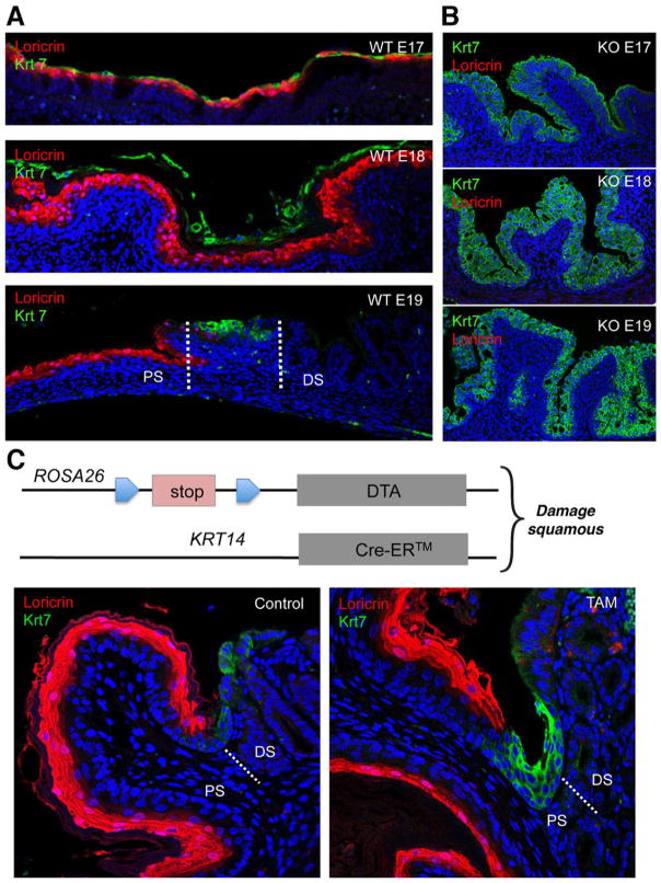
Figure 2.
Dynamics of RECs in wild-type and p63ko mice. (A) Co-labeling of squamous (anti-loricrin, red) and RECs (anti-Krt7, green) in wild-type embryos showing suprasquamous disposition of RECs at E17, sloughing of RECs at E18, and retention of RECs at the squamocolumnar junction at E19. (B) Corresponding labeling of loricrin and Krt7 in p63ko embryos showing absence of squamous cells and the development of a Barrett’s-like metaplasia in late embryogenesis. (C) Schematic of mouse strain in which diphtheria toxin A subunit is conditionally expressed in Krt14-expressing squamous stem cells. Left, section through the squamocolumnar junction staining with anti-loricrin showing squamous tissue and anti-Krt7 showing junctional RECs. Right, section through junction of mouse after 7-day expression of DTA in squamous tissue showing redistribution of Krt7 cells toward squamous epithelium. Figure 2A and B reprinted with permission from Wang et al.16 Figure C is provided with permission from Dr Xia Wang.

