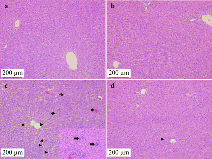
Fig. 1.
Effects of NAC administration on histopathological changes in ADR-induced liver tissue. Control (a) and NAC group (b) liver tissues had normal histological appearance. In the ADR group (c); sinusoidal dilatation (triangle), hemorrhagic areas (notched arrow), hepatocyte vacuolızation (thin arrow), foci of inflammation (thick arrow) and hemorrhagic areas (notched arrow) were observed. Histopathological changes were significantly reduced in the ADR + NAC group (d) compared to the ADR group (p < 0.05). Hematoxylin Eosin, scale bar: 200µm, × 100. ADR: Adriamycin; NAC: N-Acetylcysteine

