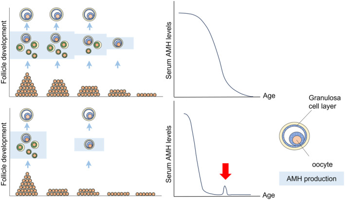
FIGURE 1.

Schematic model of the residual follicle pool and serum AMH levels; comparison of age‐dependent decline in women with normal cycles and with women POI. The upper panel shows the model for women with normal cycles: residual follicles decrease with age, accompanied by a decrease in serum AMH levels. The lower panel shows the model for women with POI: the residual follicle pool is smaller than that in age‐matched women with normal cycles. Consequently, serum AMH levels were low from an early stage of reproductive life. If there are cycles where small follicles develop, it is supposed that trace amounts of AMH can be detected in the serum (red arrow).
