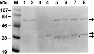
FIG. 8.
Protein profile of each reconstituted TGAH fusion protein. Lane M, molecular mass markers; lanes 1 and 3, purified Hβ and TGα subunits from plasmid pSERH; lanes 2 and 4, purified Hβ and TGα subunits from plasmid pCYSH; lane 5, reconstitution of Hβ and TGα subunits from plasmid pSERH; lane 6, reconstitution of Hβ subunit from plasmid pCYSH with TGα subunit from plasmid pSERH; lane 7, reconstitution of Hβ subunit from plasmid pSERH with TGα subunit from plasmid pCYSH; lane 8, reconstitution of Hβ and TGα subunits from plasmid pCYSH. From top to bottom, the arrows indicate the Hβ subunits from either pSERH or pCYSH, the TGα subunit from plasmid pCYSH, and the TGα subunit from plasmid pSERH, respectively. The arrowhead indicates a further processed TGα subunit from plasmid pCYSH.

