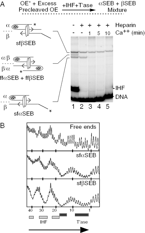
Figure 3.
Identification of two isomers of the SEB complex. (A) SEB complex was assembled by mixing a radioactively labeled uncleaved outside end with an excess of unlabeled precleaved outside end. This biases the reaction strongly in favor of the assembly of mixed complexes. The uncleaved outside end was prepared by digesting pRC167 with PstI+XhoI (96 bp transposon arm/77 bp flanking DNA). The precleaved outside end was prepared by digesting pRC35 with PvuII+BstEII (87 bp transposon arm). sfαSEB, semi-folded α-single-end-break; sfβSEB, semi-folded β-single-end-break. Other details are as given in Figures 1 and 2. (B) The sfαSEB and sfβSEB were footprinted with hydroxyl radicals. Briefly, complexes were assembled, treated in solution with hydroxyl radicals and separated using the EMSA. The complexes were recovered from the gel and the footprints were displayed on a DNA sequencing gel. Dark and light shaded boxes represent the transposase and IHF footprints as described previously (32). The large arrowhead indicates the location of the transposon end. The number of base pair inside the transposon is indicated.

