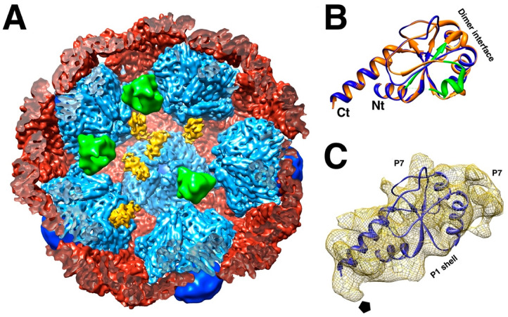
Figure 6.
The location of P7. (A) Each P7 monomer (yellow) is bound to the interface between two P1A subunits (light blue), but only at some locations around the five-fold axis. Polymerase monomers (green) are located at other sites that overlap with the P7 sites. The P1B subunits (red) and P4 hexamers (dark blue) complete the procapsid. (EMDB: 2341) (B) Homology model of the Φ6 P7 based on the crystal structure of the Φ12 P7 protein N-terminal fragment. The dimer interface in the crystal is indicated. (C) One P7 density with a fit of the homology model. Also indicated are the interface to the P1 shell and the interfaces to two potential P7 neighbors around the three-fold axis. The pentagon denotes the five-fold axis [35].

