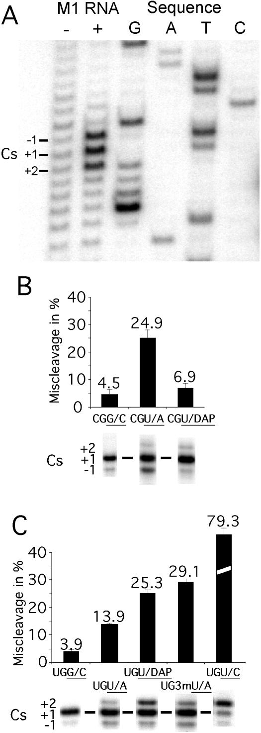
Figure 2.
Miscleavage of various substrates. (A) Identification of the cleavage site in pGlnU/A by primer extension analysis as described in Materials and Methods. In this experiment, ∼0.2 μM pGlnU/A was incubated for 8 min (after preincubation of M1 RNA for 7 min) in the presence or absence of 2 μM M1 RNA in 50 mM MES (pH 6.0) and 40 mM Mg(OAc)2. This was followed by primer extension analysis using an oligodeoxynucleotide complementary to residues A31 through C48 in tRNAGln (see Figure 1). Cs = cleavage site. (B and C) Miscleavage of various pATSer substrates [(B) with C at −1 and (C) with U at −1) represented as percentage of miscleavage and cleavage sites as indicated where for example UGG/C corresponds to the substrate pATSerUGG/C, for details see text. The 5′ cleavage fragments were separated on 22% denaturing PAGE as described in Materials and Methods.

