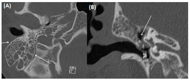
Figure 8.
CT of a 17-year-old male patient using bone windowing with axial (A) and coronal (B) views of the right middle ear. Complete obstruction of the mastoid cells is present without multiple bony sclerosis. A tympanic tube through the thickened eardrum has been placed. Obstruction of the epitympanum without bony erosion of the scutum is present.

