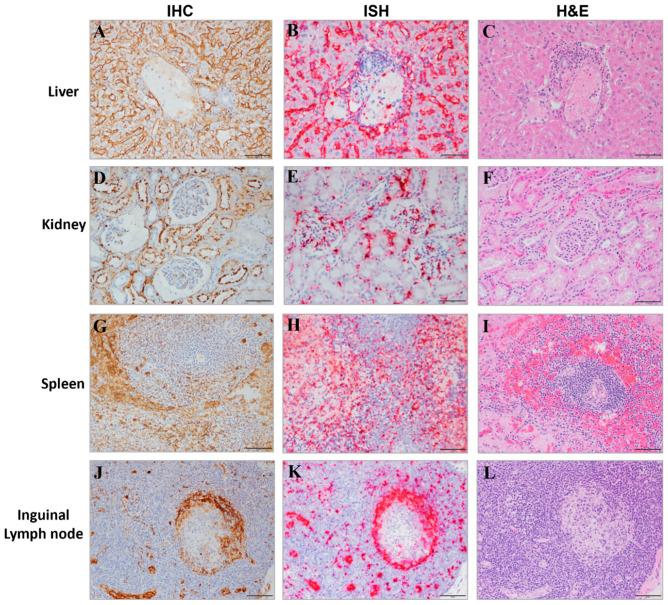
Figure 13.
Target Organs of MARV Infection. Representative images of liver (A–C), kidney (D–F), spleen (G–I), and inguinal lymph node (J–L) tissues from MARV-exposed animals are shown with IHC (left panel), ISH (middle panel) and H&E (right panel) staining from adjacent sections shown. Positive staining from IHC tissue appears as a brown precipitate and the positive ISH staining is red. The scale bar is 50 µm.

