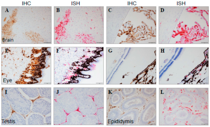
Figure 14.
Immune-Privileged Tissues from MARV-Exposed Animals. Representative images of tissues from MARV-exposed animals are shown with IHC and ISH staining from adjacent sections. Brain (A): brain parenchymal IHC, (B): adjacent brain parenchymal ISH, (C): brain ventricles/choroid plexus IHC, (D): adjacent brain ventricles/choroid plexus ISH; eye (E): IHC at low magnification, (F): IHC at high magnification, (G): ISH at low magnification, (H): ISH at high magnification; testis (I): IHC, (J): ISH; and epididymis (K): IHC, (L): ISH. Positive staining from IHC tissue appears as a brown precipitate, and positive ISH staining is red. The scale bar is 50 μm.

