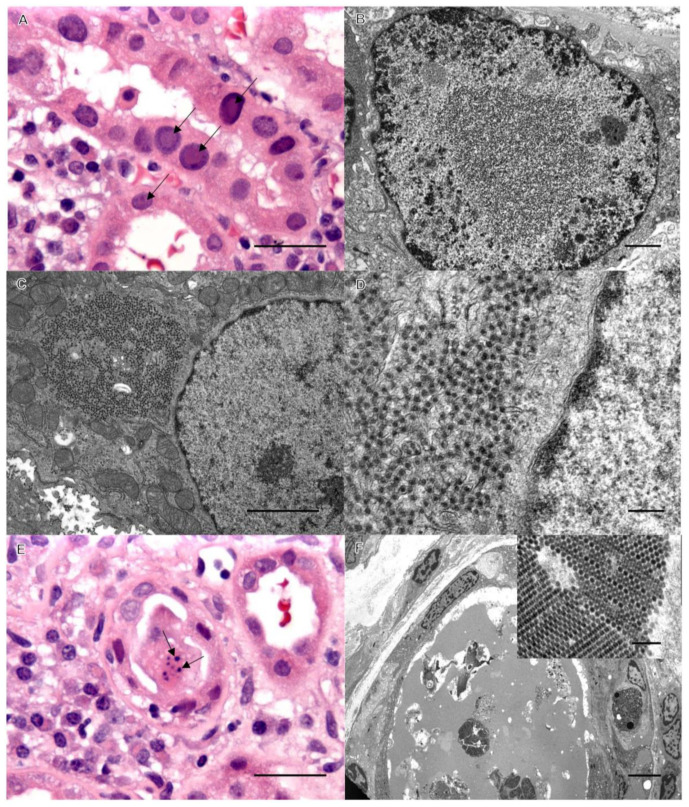
Figure 2.
Classic viral inclusions of BK polyoma virus by light microscopy in panel (A) (arrows), bar = 50 µm, and by EM in panel (B). Bar = 1 µm. Panels (C–F) demonstrate identical WNV inclusions in case 2, as in case 1. Bars: C = 1 µm, D = 0.5 1 µm, E = 50 1 µm, and F = 20 1 µm (inset bar = 200 nm).

