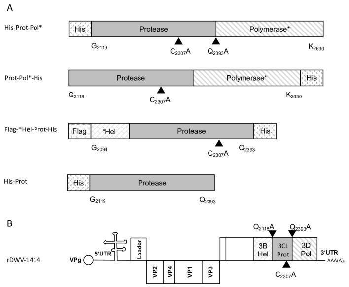
Figure 1.
Polyprotein fragments and virus genomes used in this study. (A) Scheme of the recombinant proteins used for the determination of 3CL and 3DL cleavage sites and boundaries. Protein tags (His and Flag) were attached to the N- or C-terminus of the viral polyprotein fragments as indicated. The polyprotein positions were specified according to the format of amino acid and residue number (Xn), indicating the boundaries of the recombinant polyprotein fragments. All residue numbers refer to DWV-A strain 1414. Arrowheads indicate the position of cleavage sites and the central cysteine residue of the catalytic triad. An asterisk in designations is to indicate that it is only a fragment of the respective protein. (B) Genome organization of DWV-A with annotation of the processing sites characterized in this study.

