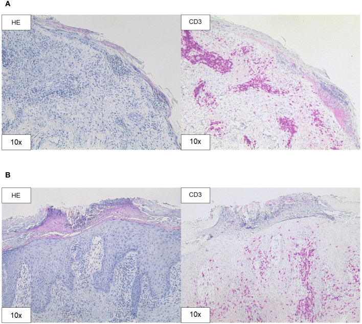
Figure 3.
Histopathology. (A) H&E and CD3 staining (original magnification 10x) of the skin biopsy of the chest demonstrating interface dermatitis with colloid bodies, perifollicular mucin, and mixed immune cells, containing lymphocytes, histiocytes, and neutrophil granulocytes. (B) H&E and CD3 staining (original magnification 10x) of the skin biopsy of the right forearm showing acanthosis, parakeratosis, intraepithelial neutrophil granulocytes, and CD3 positive T cell infiltration.

