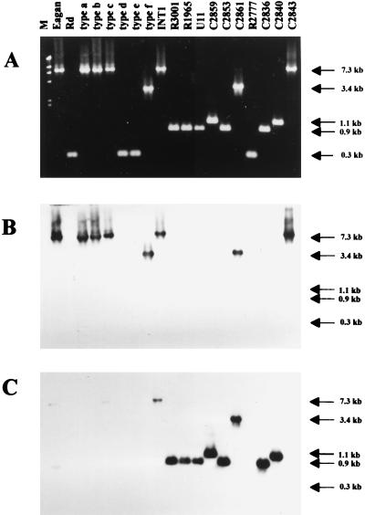
FIG. 2.
PCR amplification of the purE-pepN regions from various H. influenzae strains. (A) Genomic DNAs were amplified with primers purE-F and pepN-R (Fig. 1). Lane M, markers (λHindIII fragments). The sizes of PCR fragments, estimated from mobilities, are indicated at the right. The predicted size of the PCR fragment from Rd is 256 bp, and that from Hib AM30 is 7,270 bp. (B) The gel in panel A was blotted and probed with a hif-specific probe made by PCR with internal primers hifA-F and hifE-R. (C) The blot in panel B was stripped and reprobed with a hic probe, specific for the R3001 insert. The hic probe is a cloned, 468-bp AluI partial digestion fragment of the R3001 insert (Fig. 3).

