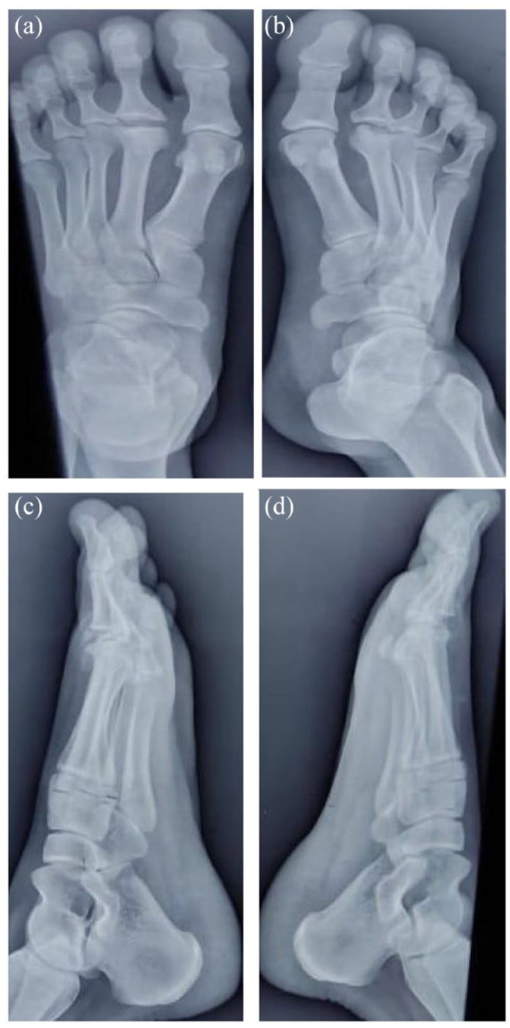
Figure 1.

X-ray foot anteroposterior (a and b) and oblique view (c and d) of the bilateral foot shows flattening of the right second metatarsal head with the widening of the joint space. Sclerosis of the articular surface is also noted. No obvious cortical deformity is seen. The rest of the metatarsal and phalangeal bones are normal. There is mild deformity noted in the left metatarsal head with flattening in the lateral surface. Increased joint space is also noted. Sclerosis is noted in the articular surface of the left second metatarsal head.
