Fig. 2.
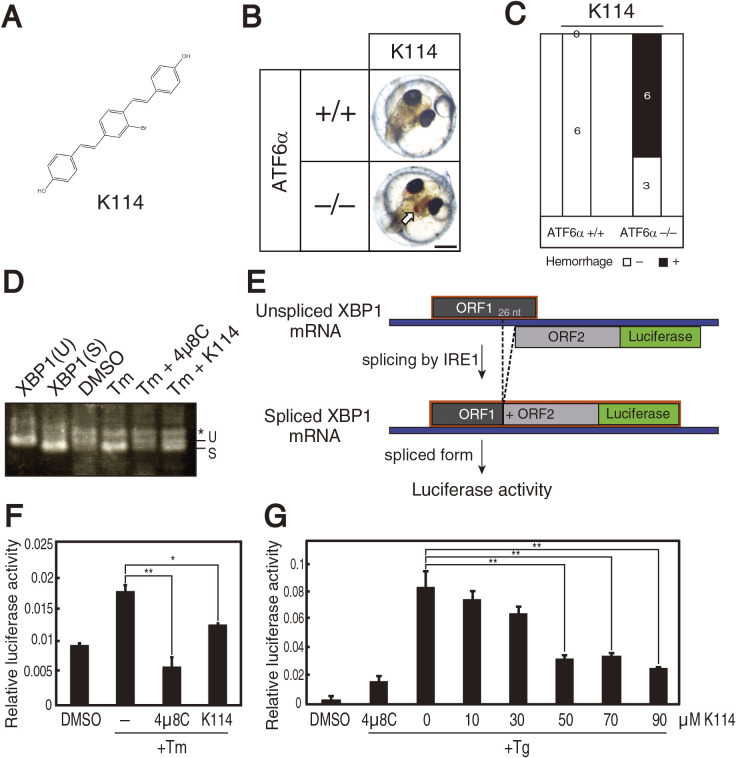
Effect of K114 on splicing of XBP1 mRNA. (A) Chemical structure of K114. (B) ATF6α+/+ and ATF6α–/– embryos at 1 dpf were treated with 30 μM K114 for 5 days, and then photographed. (C) Intracerebral hemorrhage ratio of embryos with the indicated genotypes after treatment with K114 (30 μM). (D) HCT116 cells were treated with 2 μg/ml tunicamycin with or without 4μ8C (45 μM) or K114 (50 μM) for 18 h. Total RNA were prepared and subjected to RT-PCR, as in Fig. 1D. (E) Schematic representation of the XBP1-luciderase reporter system. (F) HCT116 cells stably expressing XBP1-firefly luciferase reporter were treated with 2 μg/ml tunicamycin with or without 4μ8C (45 μM) or K114 (50 μM) for 18 h, and then cellular luciferase activity was determined. (G) HCT116 cells transiently expressing XBP1-firefly luciferase reporter were treated with 300 nM thapsigargin together with the indicated concentrations of K114 or 4μ8C (45 μM) for 18 h, and then cellular luciferase activity was determined.

