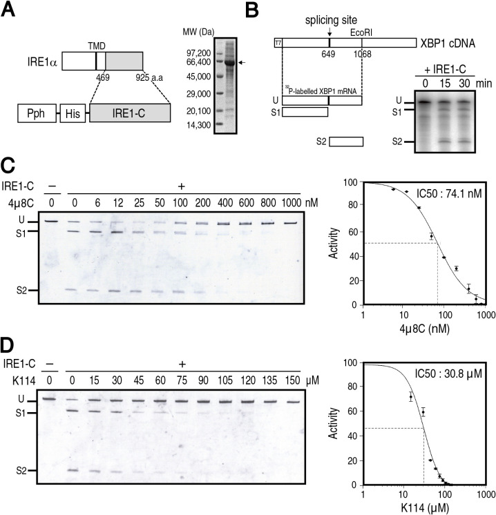
Fig. 3.
Effect of K114 on IRE1α ribonuclease activity in vitro. (A) Schematic representation of the strategy to express IRE1-C using a baculovirus system (left) and SDS-PAGE (10% gel) analysis of IRE1-C purified from SF9 insect cells (right, arrow). (B) Schematic representation of a part of XBP1 mRNA as a substrate of IRE1-C (left). Unspliced fragment (U) was cleaved to produce spliced fragments (S1 and S2). 32P-labeled XBP1 mRNA was incubated with purified IRE1-C for the indicated periods, separated by polyacrylamide gel, and autoradiographed (right). (C) (D) Unlabeled XBP1 mRNA was incubated with (+) or without (–) purified IRE1-C for 30 min in the presence of the indicated concentration of 4μ8C (C) or K114 (D), separated by urea-containing polyacrylamide gel, and stained with SyberGold (n=3). Intensity of each band was determined. Summation of S1 and S2 divided by summation of U, S1 and S2 obtained without 4μ8C or K114 is taken as 100%. The IC50 was calculated by nonlinear regression using Quest GraphTM IC50 Calculator (AAT Bioquest).

