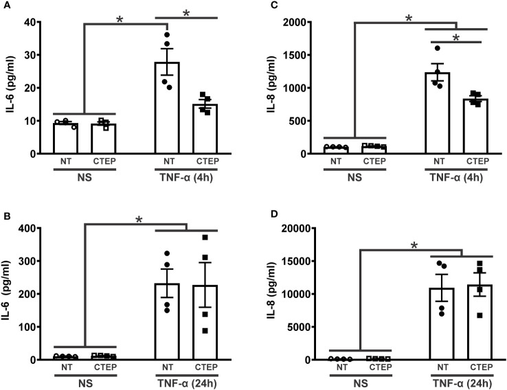
Figure 2.
CTEP treatment reduces rTNF-α-induced expression of inflammatory factors. Graphs show protein quantification of IL-6 (A) and IL-8 (C) in the supernatant of hiPSC-derived astrocytes that were either unstimulated (NS) or stimulated with rTNF-α 10 ng/mL and treated with either vehicle (NT) or CTEP 10 µM for 4 h. Graphs show protein quantification of IL-6 (B) and IL-8 (D) in the supernatant of hiPSC-derived astrocytes that were either unstimulated (NS) or stimulated with rTNF-α 10 ng/mL and treated with either vehicle (NT) or CTEP 10 µM for 24 h. Protein levels were assessed by CBA, which was performed in duplicates. Data represents the means ± SEM, n=4-6. * (p<0.05) indicates significant differences.

