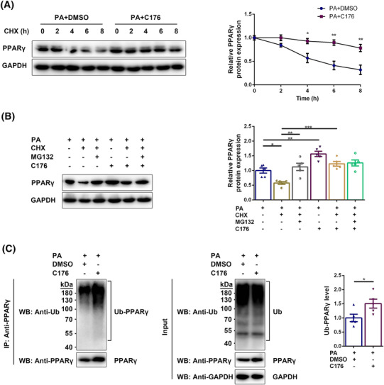
Figure 6.

STING inhibition reduced the degradation of PPARγ through the proteasome pathway in the diabetic state. Myotubes were divided into PA + DMSO group and PA + C176 group and treated with CHX for 0, 2, 4, 6, or 8 h. (A) Expression of PPARγ in myotubes detected through western blotting; GAPDH: internal reference (n = 5). Myotubes were divided into PA group, PA + CHX group, PA + CHX + MG132 group, PA + C176 group, PA + CHX + C176 group, and PA + CHX + MG132 + C176 group. (B) Expression of PPARγ in myotubes detected through western blotting; GAPDH: Internal reference (n = 5). Myotubes were divided into PA + DMSO group and PA + C176 group. Immunoprecipitation using anti‐PPARγ antibody was performed and western blotting of IP lysates and whole‐cell lysates was carried out using anti‐Ub antibody and anti‐PPARγ antibody. (C) Detection of ubiquitinated PPARγ in myotubes through western blotting; GAPDH: internal reference (n = 5). *P < 0.05, **P < 0.01, ***P < 0.001.
