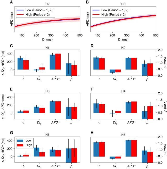
Figure 8.
The electrophysiological characteristics of areas with different periodicities. The top row (A and B) shows the overlapped restitution curves for two hearts. The curves depict regions of Periods 1 and 2 (low) and areas of Periods 3–8 (high). The restitution curves are so similar that it is difficult to separate the two curves. (C–H) The electrophysiological properties (restitution-curve parameters and conduction velocity) between each heart’s low and high periods regions. Restitution curves are parameterized by , , and τ (see the Methods section). The ratio of conduction velocity at fast pacing rates (the last three recordings before fibrillation) to the conduction velocity during slow pacing (remaining recordings) is denoted ρ.

