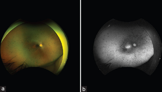
Figure 1.

(a) Wide-field retinal imaging of the right eye shows retinal pigment epithelium mottling and atrophy, scattered bone spicule pigmentation, vascular attenuation, and optic nerve pallor. The left eye (not shown) was similar; (b) Wide-field short-wave autofluorescence of the right eye shows confluent areas of hypo-autofluorescence centrally, increased hyper-autofluorescence in the central macula, and scattered areas of hypo-autofluorescence in the mid and far periphery. The left eye (not shown) was similar.
