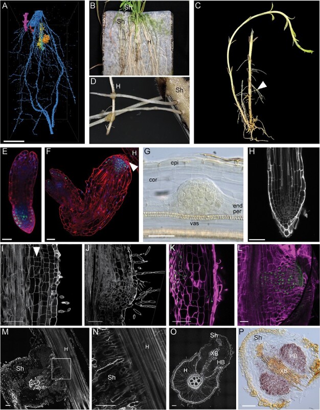
Fig. 1.

Terminal and lateral haustorium development in Striga hermonthica. (A) Micro-CT 3D reconstruction and post-processed image segmentation depicting the association of Striga seedlings (orange, yellow, and purple) attached to the rice roots (blue). (B) Macrophotograph showing Striga plants grown on rice as a host plant on top of nylon mesh. (C) Macrophotograph showing the Striga plant isolated from the host plants. Note the emerging adventitious roots (arrowhead). (D) Attachment of Striga adventitious roots to the host roots by lateral haustoria. (E and F) Confocal image showing the root meristem of Striga seedlings directly after germination (E) and before attachment to the host root (F). Roots were stained with modified pseudo-Schiff propidium iodide (mPS-PI) (red) (Truernit et al., 2008), dividing cells were visualized by 5-ethynyl-2ʹ-deoxyuridine (EdU) staining (green), and nuclei were stained with Hoechst (blue); the arrowhead points to haustorial hairs. (G) Lateral root primordium of Striga roots stained with Lugol, cleared with chloral hydrate, and visualized with Nomarski microscopy. (H) Confocal image of a Striga lateral root tip; cell walls stained with mPS-PI. (I and J) Confocal images of longitudinal vibratome sections of a Striga root developing a lateral haustorium; cell walls and starch granules were stained with mPS-PI; the arrowhead points to periclinal cell divisions in the inner cortex upon haustorium initiation. (K and L) Confocal longitudinal sections showing cell divisions in developing Striga lateral haustoria. EdU-stained nuclei are green and cell walls stained with SCRI Renaissance are in magenta. (M) Longitudinal section of a Striga lateral haustorium during attachment to the host plant; (N) magnification of the area marked in (M). (O) Cross-sections of a Striga lateral haustorium attached to a host root; cell walls stained with mPS-PI. (P) Starch granules accumulation in the Striga lateral haustorium, visualized by Lugol’s staining. cor, cortex; end, endodermis; epi, epidermis; H, host; HB, hyaline body; per, pericycle; Sh, Striga hermonthica; vas, vasculature; XB, xylem bridge. Scale bars 20 μm (A, B), 50 μm (E, F, G, H, I, J, K, L, M, N, O, P).
