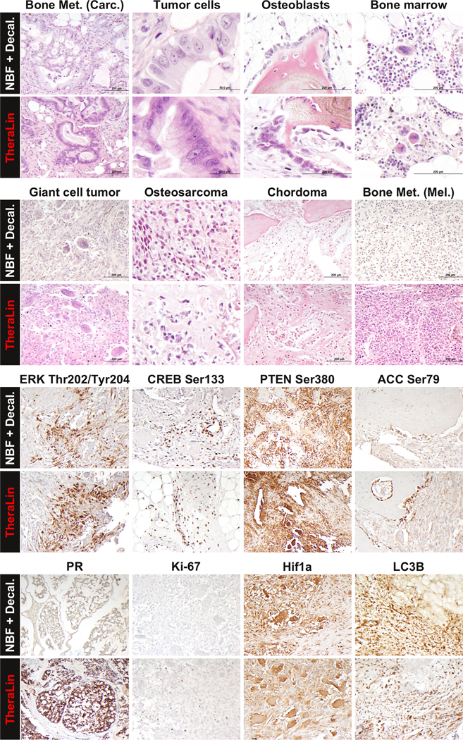
Figure 3. Representative images comparing tissue morphology and IHC stains of Theralin fixed and formalin fixed, decalcified bone tumor samples.

Patient matched tissue sections were stained with either Hematoxylin and eosin (top half) or for selected IHC targets (bottom half).
