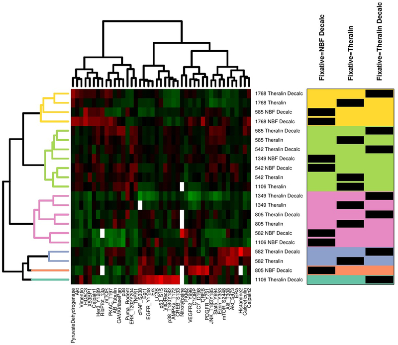
Figure 6. Formalin fixed and decalcified samples cluster away from patient matched Theralin fixed and Theralin fixed and decalcified samples during 2-way unsupervised hierarchical clustering.

Tumor cells from seven patient matched samples that were either Theralin fixed, Theralin fixed and decalcified, or formalin fixed and decalcified were laser-capture microdissected and lysates printed on reverse phase protein microarrays. Relative protein levels were normalized to the total extracted protein content per sample due to the reduced protein extractability from formalin fixed samples. Numbers in center column represent patient IDs. Box on right shows fixation type per sample (black bars) within each cluster (color shaded areas). (Decalc = decalcified, NBF = formalin fixed)
