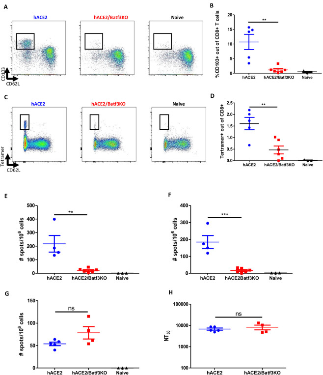
Fig 2. The cDC1 subset is required for induction of CD8 memory T-cells following SARS-CoV-2 infection.
hACE2/Batf3KO (red) and hACE2 (blue) mice were infected i.n with 50pfu of SARS-CoV-2. Results obtained with naïve non-infected mice are also depicted (black). 7 weeks post infection the frequencies of mucosal imprinted CD103+ memory CD8 T-cells was evaluated (A-representative FACS analysis from each group and B- histograms incorporating the individual sets of results obtained for each animal). Memory CD8 T-cells specific for SARS-CoV-2 in the lung were quantified by tetramer staining (C and D, as above). ELIspot assays were performed using splenocytes from infected animals stimulated with the S539 (E) and S915 (F) peptides. SARS-CoV-2 specific CD4 T-cells were quantified on splenocytes from infected animals using ORF3 peptide (G). Neutralizing antibody titers were determined by pseudovirus neutralization assay (H). Bars indicate means ± SEM from 4–6 animals per group. Gating strategy for A-D appear in S7 Fig. P values: *P < 0.05, **P < 0.01, ***P < 0.001, and ns, not significant.

