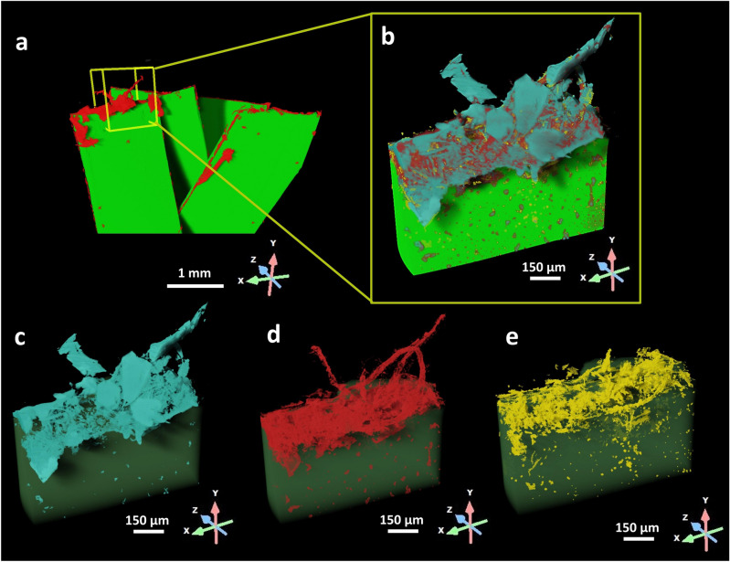
Figure 3.
3D model reconstruction and phase segmentation. The high resolution XRM dataset corresponding to the edge colonisation growth site was further examined to investigate the composition of the sample. (a) 3D model of PET fragments (green) colonised by F2 F. oxysporum (red) where the yellow box highlights the VOI scanned using high-resolution XRM. (b) The VOI scanned in high resolution mode where three different phases were segmented: (c) salts crystals in light blue, (d) the fungus in red and (e) the biodegraded plastic due to the fungal strain attack in yellow.

